Location
3/4, Sarswati Plaza Opp. N.A.S. College, E.K. Road, Meerut

Dr. Vineet Bansal's Clinic, located at 3/4, Saraswati Plaza, Opposite N.A.S. College, E.K. Road, Meerut, is renowned for its excellence in cardiology. With exceptional expertise, Dr. Vineet Bansal is committed to delivering high-quality, personalized healthcare services.
 By Dr. Vineet Bansal
 Sept 16, 2026
The heart is one of the most important organs in the human body, continuously pumping blood to deliver oxygen and nutrients throughout the body. Understanding how well the heart functions is essential for identifying certain cardiovascular conditions and guiding appropriate medical care.
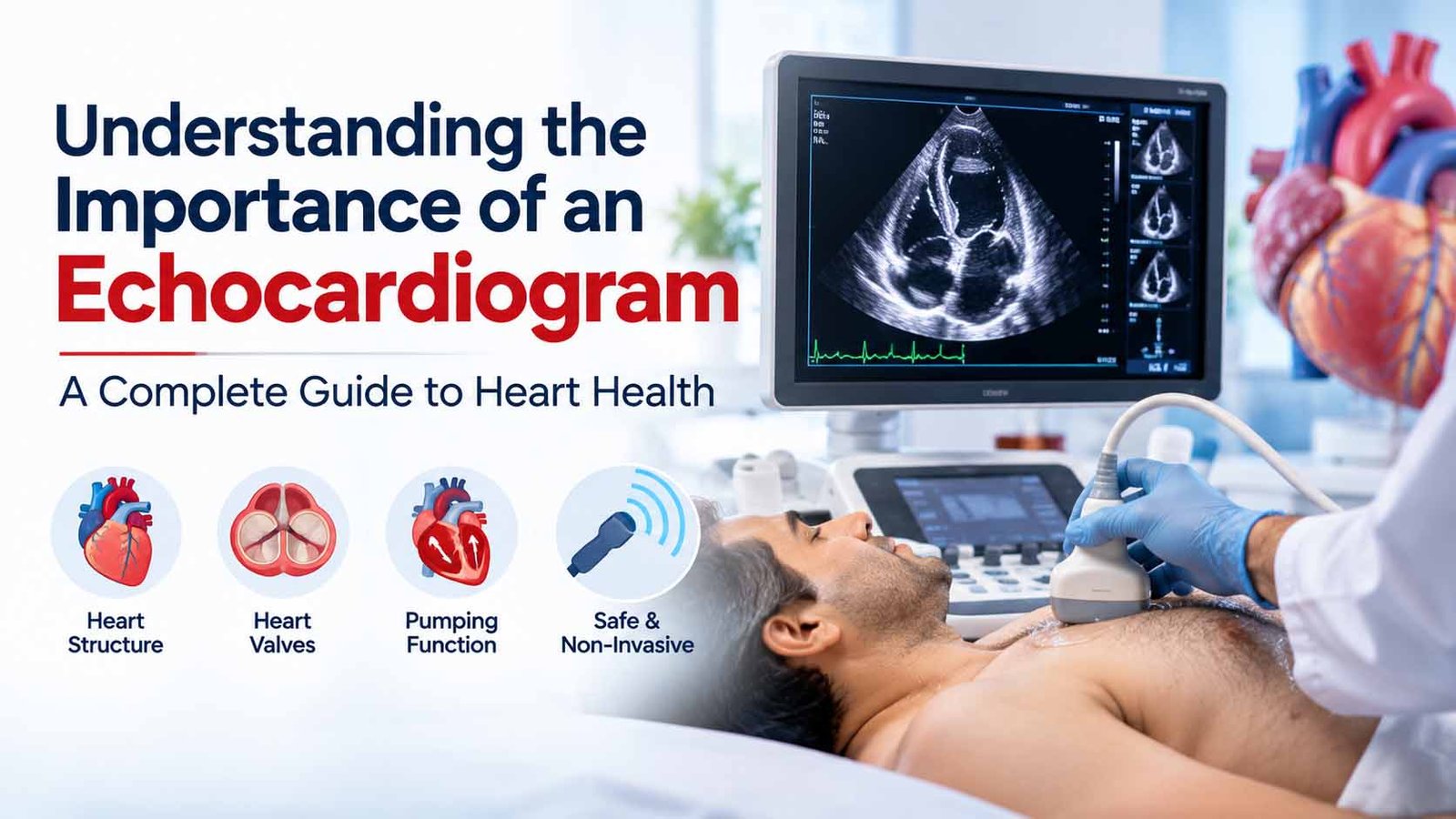
An echocardiogram, commonly known as a 2D Echo or heart ultrasound, is a widely used diagnostic test that allows healthcare professionals to examine the structure and function of the heart without surgery.
This imaging technique helps cardiologists assess the heart's chambers, valves, pumping ability, and blood flow patterns.
Dr. Vineet Bansal, MBBS, MD, DM (Cardiology), a cardiologist in Meerut, emphasizes the importance of understanding cardiac diagnostic tests and their role in evaluating heart health.
An echocardiogram is a medical imaging test that uses high-frequency sound waves to produce moving images of the heart.
During the test, a device called a transducer sends ultrasound waves through the chest. These sound waves reflect off the heart's structures and are converted into images displayed on a monitor.
An echocardiogram can provide information about:
Unlike X-rays or CT scans, standard echocardiography does not use ionizing radiation.
An echocardiogram plays an important role in diagnosing and monitoring many cardiovascular conditions.
Some heart conditions can affect the structure or function of the heart before severe symptoms develop.
Echocardiography provides useful information that helps healthcare professionals understand these changes and determine whether further investigations or treatment may be necessary.
One of the most important uses of an echocardiogram is assessing how effectively the heart pumps blood.
A commonly reported measurement is the left ventricular ejection fraction (LVEF), which estimates the percentage of blood pumped out of the left ventricle with each heartbeat.
An echocardiogram can also evaluate other aspects of heart muscle function.
These findings help assess conditions such as heart failure and certain cardiomyopathies.
The heart contains four valves that help maintain blood flow in the correct direction.
An echocardiogram can help identify conditions such as:
The test helps assess whether a valve is narrowed, leaking, or functioning abnormally.
Some cardiovascular conditions cause the heart muscle to become thickened, enlarged, weakened, or stiff.
An echocardiogram can help identify structural and functional changes associated with:
These findings can support diagnosis and ongoing medical management.
Congenital heart disease refers to structural abnormalities of the heart that are present from birth.
An echocardiogram can help identify some congenital conditions, including:
Depending on the suspected condition, specialized echocardiography or additional imaging may be required.
An echocardiogram is not only used to investigate new symptoms. It can also help monitor individuals with previously diagnosed cardiovascular conditions.
For example, repeat echocardiography may be appropriate for selected patients with:
The need and frequency of repeat testing depend on the patient's condition and clinical recommendations.
Different echocardiography techniques are available depending on the clinical information required.
This is the most common type of echocardiogram.
A healthcare professional places an ultrasound transducer on the chest to obtain images of the heart.
It is generally painless, non-invasive, and does not require anesthesia.
Doppler ultrasound evaluates the direction and speed of blood flow through the heart and its valves.
It is commonly used alongside standard echocardiography to assess valve abnormalities and certain pressure-related findings.
A transesophageal echocardiogram uses a specialized ultrasound probe passed into the esophagus to obtain detailed images of certain heart structures.
It may be recommended when standard chest-based imaging does not provide sufficient information.
Unlike a routine transthoracic echo, TEE is semi-invasive and generally involves specific preparation, monitoring, and often sedation.
A stress echocardiogram evaluates how the heart functions during physical exercise or medication-induced stress.
It may be used in selected patients to investigate suspected coronary artery disease or assess certain other cardiac conditions.
Stress echocardiography is not routinely necessary for everyone.
3D echocardiography provides additional structural detail and may be useful in selected cases, particularly for evaluating heart valves and complex cardiac anatomy.
A cardiologist or another qualified healthcare professional may recommend an echocardiogram when symptoms or examination findings suggest a possible structural or functional heart problem.
Common reasons include:
An echocardiogram may also be recommended in specific situations involving suspected congenital heart disease or other structural abnormalities.
However, not every person with chest pain, palpitations, or high blood pressure requires an echocardiogram.
The decision should be based on medical history, physical examination, and individual clinical findings.
A standard transthoracic echocardiogram is usually a straightforward outpatient procedure.
The patient is asked to lie on an examination table. Electrodes may be placed on the chest to monitor the heart's electrical activity during imaging.
A water-based gel is applied to the chest to help transmit ultrasound waves.
A trained professional moves the ultrasound transducer across different areas of the chest to obtain images of the heart.
The patient may be asked to change position or briefly hold their breath to improve image quality.
The images are reviewed and interpreted by an appropriately qualified healthcare professional.
The report describes relevant findings, such as heart chamber dimensions, pumping function, valve function, and other structural observations.
A routine transthoracic echocardiogram commonly takes approximately 30–60 minutes, although the duration varies depending on the examination.
A standard transthoracic echocardiogram is generally considered a safe diagnostic test.
It uses ultrasound rather than ionizing radiation and does not usually require injections or anesthesia.
Some patients may experience mild temporary discomfort from pressure applied by the transducer.
Other types of echocardiography, such as TEE and stress echocardiography, involve additional considerations and potential risks that should be discussed with the healthcare team.
This is a common question among patients.
A standard resting echocardiogram cannot directly visualize most blockages in the coronary arteries.
However, it may identify abnormalities in heart muscle movement or pumping function that can sometimes be associated with reduced blood supply or previous heart muscle damage.
A normal resting echocardiogram does not rule out coronary artery disease.
If coronary artery disease is suspected, a healthcare professional may recommend other investigations, such as stress testing, coronary CT angiography, or invasive coronary angiography, depending on the clinical situation.
An echocardiogram report may include several medical terms and measurements.
Some commonly reported findings include:
Ejection Fraction (EF): An estimate of how much blood the left ventricle pumps out with each contraction.
Left Ventricular Function: Describes the pumping performance and other functional characteristics of the left ventricle.
Valve Function: Indicates whether the heart valves open and close appropriately.
Chamber Dimensions: Describes the size of different heart chambers.
Wall Motion: Evaluates how different areas of the heart muscle contract.
Pericardial Effusion: Refers to fluid accumulation around the heart.
Results should be interpreted alongside symptoms, clinical examination, and other investigations.
An isolated measurement should not be used to diagnose or exclude a heart condition without appropriate medical assessment.
Echocardiography can be valuable when a person's symptoms, examination findings, or medical history suggest a possible heart condition.
It may help identify structural or functional abnormalities and support timely clinical decisions.
However, routine echocardiography is not recommended as a universal screening test for all healthy adults without symptoms.
Preventive cardiovascular care primarily focuses on assessing and managing established risk factors, including high blood pressure, elevated cholesterol, diabetes, smoking, physical inactivity, and family history.
A qualified healthcare professional can determine whether echocardiography would provide useful additional information.
Understanding cardiac diagnostic tests can help individuals make informed decisions about their cardiovascular health.
For patients in Meerut and surrounding areas, awareness of the purpose, benefits, and limitations of an echocardiogram is important.
Dr. Vineet Bansal, MBBS, MD, DM (Cardiology), is a cardiologist based in Meerut, Uttar Pradesh.
Understanding when echocardiography is appropriate can help patients discuss their symptoms, diagnostic findings, and heart health concerns with their healthcare professionals.
An echocardiogram is used to evaluate the structure and function of the heart, including heart chambers, valves, pumping ability, and blood flow patterns.
A routine transthoracic 2D Echo is generally painless. Mild discomfort may occur when the ultrasound transducer is pressed against the chest.
A routine transthoracic echocardiogram usually takes approximately 30–60 minutes, although the duration can vary.
An echocardiogram may identify changes in heart muscle movement or function associated with a heart attack. However, it cannot independently confirm or rule out a heart attack. Clinical assessment, ECG, and blood tests are often essential.
Neither test is universally better. An ECG records the heart's electrical activity, while an echocardiogram evaluates its structure and function. They serve different purposes.
No. A normal echocardiogram is reassuring for the structures and functions assessed, but it does not exclude all cardiovascular diseases, including coronary artery disease or intermittent rhythm abnormalities.
Not necessarily. The need for repeat echocardiography depends on symptoms, existing heart conditions, previous findings, and medical recommendations.
An echocardiogram is an important diagnostic tool that helps healthcare professionals understand the structure and function of the heart.
From evaluating heart valves and pumping function to identifying certain congenital abnormalities and monitoring existing cardiovascular conditions, echocardiography provides valuable clinical information.
Understanding the benefits and limitations of an echocardiogram can help patients make informed healthcare decisions.
The test should be recommended according to individual medical needs rather than performed routinely without a clear clinical reason.
Medical Disclaimer: This article is intended solely for public awareness and educational purposes. It does not constitute personalized medical advice, diagnosis, or treatment. Diagnostic investigations should be undertaken based on appropriate medical evaluation. Seek emergency medical assistance for severe chest pain, sudden breathlessness, fainting, or other symptoms suggesting a cardiac emergency.